50 formamide Product No. A powerful tool for mRNA detection Abstract.

D8906 01 SDS Product No.

Fluorescence in situ hybridization probes. These probes are FISH confirmed on normal peripheral blood in both interphase nuclei and metaphase spreads. Article in Russian Bogomolov AG Karamysheva TV Rubtsov NB. Sensitive enough to detect individual molecules of mRNA Stellaris FISH probe.
Translocations will result in splitting of the two colors while. Large insert genomic DNA clones such as bacterial artificial chromosome BAC clones and repetitive DNA sequences have been the most commonly used DNA probes for FISH. L4390 05-15 ngµl labeled probe and 300 ngml Salmon Sperm DNA Product No.
Anzeige Used In Immunocytochemistry Immunohistochemistry and in situ Hybridization Applications. Enhance Signal and Detection Capabilities for Low-Abundance Targets. Developed by scientists for scientists OGT understands the real.
Gene specific probes are available in both single and dual colors. A new fluorescence in situ hybridization FISH method that uses peptide nucleic acid PNA probes for identification of Candida albicans directly from positive-blood-culture bottles in which yeast was observed by Gram staining herein referred to as yeast-positive blood culture bottles is described. Using spectrally distinct fluorophore labels for each hybridization probe this approach gives you the power to resolve several genetic elements or multiple gene expression patterns through multicolor visual display.
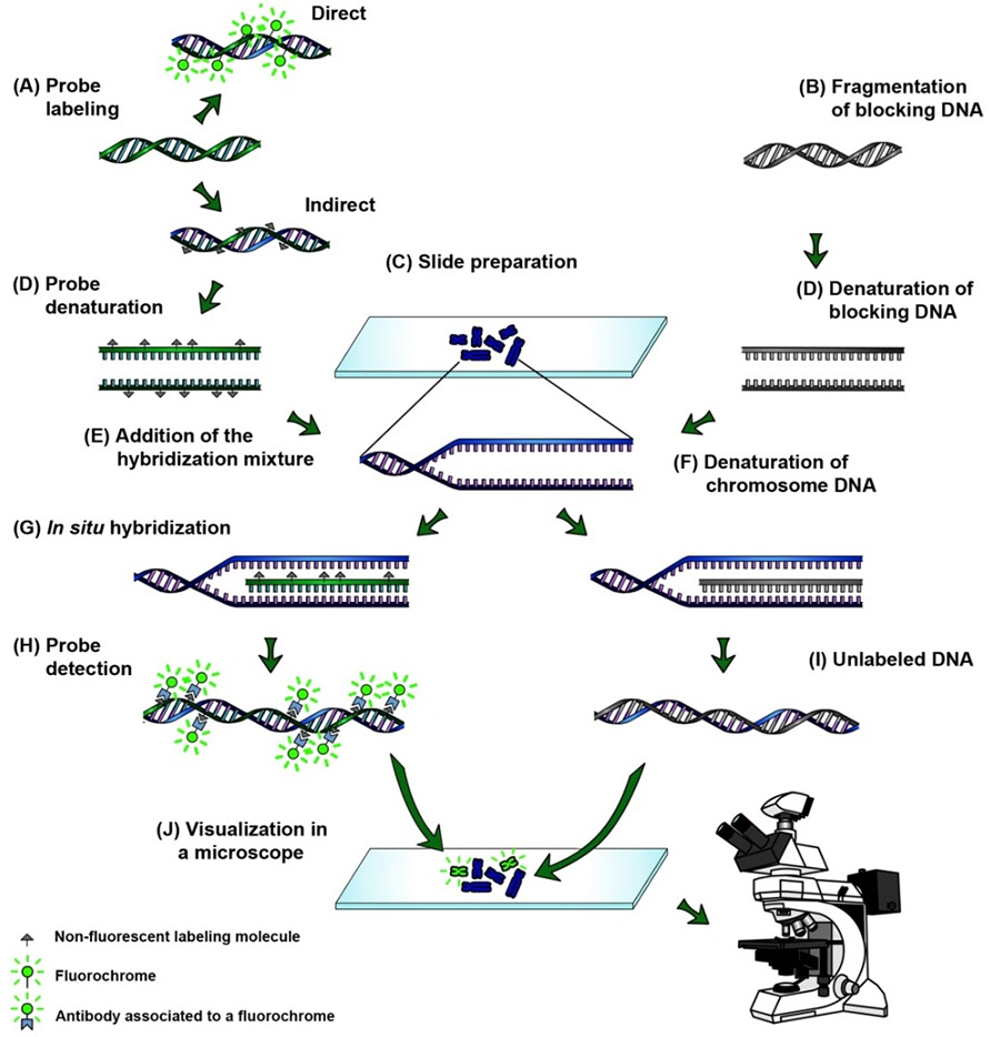
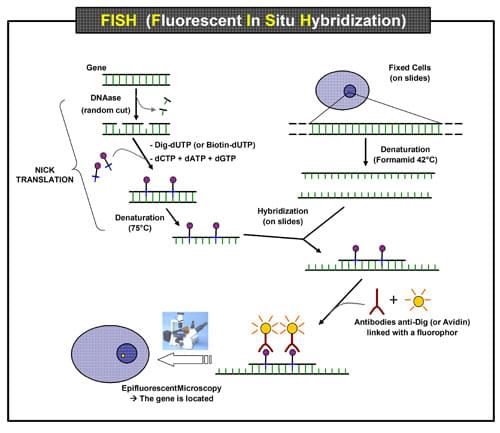
Abnova provides over 600 FISH probes for identification of gene amplification split. In this chapter we provide a systematic overview of the published guidelines and validation procedures for fluorescence in situ hybridization FISH probes for clinical diagnostic use. Plasmid DNA is labeled with biotin-11-dUTP using nick translation random priming or the polymerase chain reaction eg ADVANCE Nick Translation Kit.
D7656 in 2x. 4 Zeilen What is Fluorescent in situ hybridization. Fluorescent in situ hybridization FISH has been the most popular technique for chromosome identification in plants.
Enhance Signal and Detection Capabilities for Low-Abundance Targets. In the FISH The nucleotide sequence can be mapped. Stellaris fluorescence in situ hybridization FISH probes.
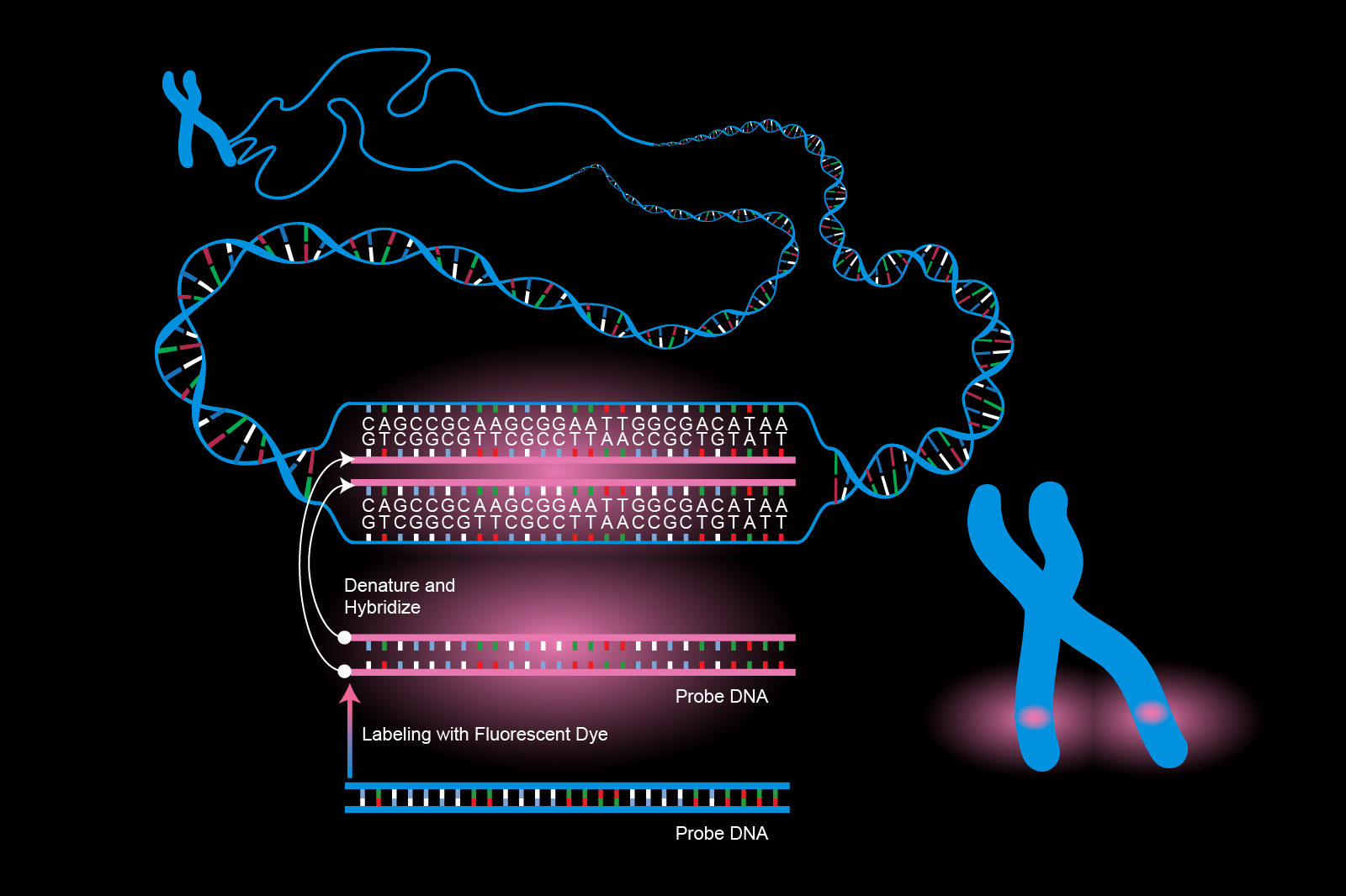
This technique exploits the use of fluorescent dye-labeled probes against a nucleotide sequence of DNA that is mapped or localized to a chromosome or intra-nuclear DNA to generate a fluorescence signal which in turn is visualized in situ by microscopy. Our dual color FISH probes allow for the detection of two genetic loci in a single sample with a different color dye assigned to each probe. Recently FISH has been extended with.
Our fluorescence in situ hybridization FISH assay also demonstrated that these probes could be used as cross-species markers between or within the genera of Sorghum and Saccharum. Fluorescence in situ hybridization with DNA probes derived from individual chromosomes and chromosome regions. Multiplex fluorescence in situ hybridization FISH enables you to assay multiple targets and visualize colocalized signals in a single specimen.
Gene Specific FISH Probes. Anzeige Used In Immunocytochemistry Immunohistochemistry and in situ Hybridization Applications. The Stellaris FISH technology is a new mRNA detection method that enables simultaneous detection.
FISH probes-which are classified as molecular probes or analyte-specific reagents ASRs-have been extensively used in vitro for both clinical diagnosis and research. This is useful for cases where multiple genes or regions need to be identified including when testing for gene translocations and fusions. Fluorescent in situ hybridization FISH Fluorescent in situ hybridization has become a well-known method for genetic mapping and gene expression profiling.
ASR and CE marked gene specific probes are available. A significant part of the eukaryotic genomes consists of repetitive DNA which can form large clusters or distributed along euchromatic chromosome regions. Gene Specific probes are designed to hybridize to a precise gene location.
Fluorescence In Situ Hybridization FISH Preparation of FISH probe Recommended Filter Set FISH is a technique used to identify and localize the presence or absence of specific DNA sequences on cells and tissues. F7508 10 dextran sulfate Product No. Fluorescence in situ hybridization FISH can be used to visualize spatial locations of DNA regions and RNA molecules in a sequence-specific manner.
High quality reliable and easy-to-use DNA probes for fluorescence in situ hybridisation FISH Highly specific CytoCell FISH probes are capable of detecting genetic changes in a variety of sample types in situ.
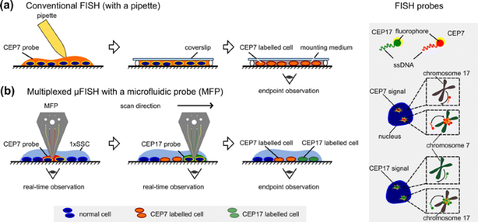 Micro Fluorescence In Situ Hybridization Mfish For Spatially Multiplexed Analysis Of A Cell Monolayer Springerlink
 In Situ Hybridization Ish And Fluorescence In Situ Hybridization Fish Creative Diagnostics
Https Biomedizin Unibas Ch Fileadmin User Upload Biomedizin Core Facilities Histology Pictures Seminars In Histology Seminar 3 R Pdf
 Fluorescent In Situ Hybridization Fish Assay Youtube
 Fluorescence In Situ Hybridization Fish Learn Science At Scitable
 A Technical Review And Guide To Rna Fluorescence In Situ Hybridization Peerj
 In Situ Hybridization Technique To Detect Mrna Localization Youtube
 Preparation Of Fluorescent In Situ Hybridisation Probes Without The Need For Optimisation Of Fragmentation Sciencedirect

 Fluorescence In Situ Hybridization Fish Creative Diagnostics
 Genevogue Biotechnology Fluorescence In Situ Hybridization Fish
 Fluorescence In Situ Hybridization Fish
 Fluorescence In Situ Hybridization Fish With Probes For Psc200 Red Download Scientific Diagram
 Fluorescent In Situ Hybridization Fish Creative Biolabs
Comments
Post a Comment